Non Peer-Reviewed Preprint
3. Q Duan, S Okuwa, R Estrella, C Yeung, YCD Chen, LQ Rio, KM Vien, and PC Volkan. Deciphering the combinatorial expression pattern and genetic regulatory mechanisms of Beats and Sides in the olfactory circuits. BioRxiv, June 3, 2025 (available online). doi: 10.1101/2025.05.31.657193. [pdf]
2. HP Gupta, A Azevedo, YCD Chen, K Xing, P A Sims, E Varol, and R Mann. Decoding neuronal wiring by joint inference of cell identity and synaptic connectivity. BioRxiv, March 4, 2025 (available online). doi: 10.1101/2025.03.04.640006. [pdf]
1. I Holguera, YC Chen, YCD Chen, F Simon, AG Gaffney, JD Rodas, S Cordoba, and C Desplan. Temporal and Notch identity determine layer targeting and synapse location of medulla neurons. BioRxiv, January 6, 2025 (available online). doi: 10.1101/2025.01.06.631439. [pdf]
Peer-Reviewed Publications
Peer-Reviewed Research and Review Articles
15. J Walsh, IP Junker, YCD Chen, YC Chen, H Gifford, DS Chen, and Y Ding. High-resolution comparative single-cell transcriptomics of doublesex-expressing neurons reveals evolutionary conservation and diversity of sexual circuits in Drosophila. PNAS, November 17, 2025;122 (47) e2516083122. doi:10.1073/pnas.2516083122. [Link] [pdf] [scMarco for dsx dataset]
A central challenge in evolutionary neuroscience is understanding how neural circuits diversify to produce species-specific behaviors while retaining conserved architecture. This question is especially relevant in Drosophila, where closely related species display striking differences in male courtship despite sharing similar brain structures and genetic programs. Sexual circuits labeled by doublesex (dsx⁺) neurons provide a powerful model for addressing this gap, yet the field has lacked a high-resolution, cross-species comparison of their cellular composition and molecular identities. We set out to determine which aspects of dsx⁺ circuits are evolutionarily conserved, which features diverge, and how these changes might underlie behavioral diversification.
Using high-resolution single-cell transcriptomics across four Drosophila species, the study identifies 84 distinct dsx⁺ neuron types and reveals that the overall cellular architecture of sexual circuits is deeply conserved, with minimal gain or loss of cell types over evolution. However, transcriptomic comparisons uncover extensive heterogeneity in how these conserved neuron types diverge. Core identity features—such as fruitless expression, neurotransmitter and monoamine usage, and key transcription factors—remain stable, whereas neuropeptide signaling pathways show dramatic, cell-type-specific evolutionary turnover. These results suggest that behavioral diversification arises not from reshaping circuit structure but from fine-scale molecular remodeling within conserved cell types. We further show that male-specific neurons are not disproportionately divergent and provide an online resource for exploring cell-type-specific markers, establishing a foundational framework for studying the evolutionary basis of behavioral adaptation.
14. JHM Soffers, E Beck, D Sytowski, M Maughan, D Devasi, Y Zhu, B Wilson, YCD Chen, T Erclik, JW Truman, JB Skeath, and H Lacin. A library of lineage-specific driver lines connects developing neuronal circuits to behavior in the Drosophila Ventral Nerve Cord. eLife, June 10, 2025; 14:RP106042. do: 10.7554/eLife.106042. doi:10.7554/eLife.106042. [Link] [pdf]
A major gap in developmental neuroscience has been the lack of genetic tools that can specifically and stably label distinct neuronal lineages throughout development and into adulthood. This limitation has made it difficult to study how neurons assemble into functional circuits and contribute to behavior over time, especially in complex nervous systems like that of the fruit fly, Drosophila melanogaster.
This study fills that gap by creating a large library of split-GAL4 driver lines that target individual hemilineages—developmentally related groups of neurons—in the fly’s ventral nerve cord. These genetically engineered lines remain active across developmental stages, allowing us to visualize how neuronal morphology changes, identify neurotransmitter profiles, and activate specific lineages using optogenetics. By linking each hemilineage to particular behaviors, we provide a powerful framework for exploring how neural circuits are built and function from development to adulthood.
13. Y Carrier#, LQ Rio#, N Formicola, V de Sousa-Xavier, M Tabet, YCD Chen, AH Ali, M Wislez, L Orts, A Borst, and F Pinto-Teixeira. Biased cell adhesion organizes the Drosophila visual motion integration circuit. Developmental Cell, November 15, 2024 (available online). doi: 10.1016/j.devcel.2024.10.019. [Link] [pdf] #Co-first authors
In the brain, different layers of neurons connect with specific partners to perform precise functions. In the visual system of fruit flies, certain neurons called T4 and T5 specialize in detecting motion and send their signals to specific layers where they connect with other neurons. In this collaborative work with Pinto-Teixeira’s group, we discovered that cell adhesion molecules called Beat and Side, which help cells stick together, play a key role in ensuring these neurons connect in the correct layers. By studying how these proteins work during development, we found that they do not directly create the connections between neurons. Instead, they help neurons group into the right layers, ensuring they are close enough to form the correct connections later. This process creates highly organized and accurate wiring in the brain, allowing neurons to communicate with the right partners and perform their specialized roles.
12. SA Li#, HG Li#, N Shoji, C Desplan, and YCD Chen*. Protocol for replacing coding intronic MiMIC and CRIMIC lines with T2A-split-GAL4 lines in Drosophila using genetic crosses. Star Protocols, December 15, 2023; (4) 102706. doi: 10.1016/j.xpro.2023.102706. [Link] [pdf] [Media] #Co-first authors; *Corresponding author
This protocol provides a step-by-step procedure for generating gene-specific split-GAL4 drivers from coding intronic MiMIC/CRIMIC lines in Drosophila. It offers a cost-effective alternative to traditional microinjection methods for converting coding intronic MiMIC/CRIMIC lines into gene-specific split-GAL4 lines that are adaptable for fly researchers working on different tissues.
11. YCD Chen*, YC Chen, R Rajesh, N Shoji, M Jacy, H Lacin, T Erclik, and C Desplan*. Using single-cell RNA sequencing to generate predictive cell-type-specific split-GAL4 reagents throughout development.
PNAS, July 31, 2023;120 (32) e2307451120. doi: 10.1073/pnas.2307451120. [Link] [pdf] [Media] [Kudos by PNAS] [PNAS Commentary by Liqun Luo’s group]*Co-corresponding authors
In Drosophila, the availability of cell-type-specific so-called drivers for selectively expressing genes of interest in a cell-type-specific way has revolutionized the study of circuit function and behavior. These reagents largely come from a comprehensive screen of small fragments of DNA taken from genes expressed in neurons. These typically give rise to expression domains encompassing many cell types. By intersecting two lines and utilizing the split transcriptional DNA binding and transcription activation domains of GAL4 expressed in each of these lines allows for often highly specific expression in specific cell types. These so-called Split-GAL4 lines have been particularly important in providing highly reliable cell-type-specific reagents for manipulating adult neural circuitry in prescribed ways. In principle, a similar approach could be taken for studying development. Regrettably, the adult expression patterns driven by specific DNA fragments can be quite different than patterns during development. It would be ideal to have a way to reliably develop cell-type specific drivers for developmental studies. Here we describe a clever and powerful way to accomplish this.
We leverage the extensive single-cell sequencing data available for the developing fly visual system to identify specific genes and combinations of genes expressed only in specific cell types. We show that by computationally mining these data, one can identify specific genes and combinations of two of them selective for a single cell type at multiple developmental stages for the majority of cell types in the visual system. We demonstrate that these can drive cell-type-specific expression patterns during development as predicted in populations of specific neuron types or in a sparsely labeled fashion to support analysis of cellular morphology at the single-cell level. These reagents and the approach pioneered here provide extraordinary reagents for the field to ask developmental questions in a particularly precise way. Although we have focused on the fly visual system, it is clear that this approach can be used in any region of Drosophila where single cell developmental transcriptomic analysis is available.
10. YCD Chen, V Menon, RM Joseph, and A Dahanukar. Control of sugar and amino acid feeding via pharyngeal taste neurons. Early release in The Journal of Neuroscience, July 7, 2021;41 (27) 5791-5808. doi: 10.1523/JNEUROSCI.1794-20.2021. [Link] [pdf] [Media]
Drosophila has multiple taste organs throughout the body to detect chemicals that signal palatability or noxious quality. It is still unclear how individual neurons in each taste organ act to control feeding. Here, we use the fly pharynx as a model to study the extent to which taste information is integrated at the cellular level to regulate the consumption of sugars and amino acids. We generate a taste-blind fly and establish a critical role of pharyngeal input in food selection. We also study the feeding behavior of flies in which only selected pharyngeal neurons are restored and show examples of taste integration as well as redundancy in how pharyngeal neurons control the overall consumption of sugars and amino acids.
9. BC Choi, YCD Chen and C Desplan. Building a circuit through correlated spontaneous neuronal activity in the developing vertebrate and invertebrate visual systems. Genes & Development, May 1, 2021;35(9-10):677-691. doi: 10.1101/gad.348241.121. [Link] [pdf]
Understanding the role of spontaneous activity in the developing nervous system has been advancing rapidly due to a wealth of sophisticated genetic and optogenetic tools and imaging techniques. In this review, we present the current knowledge of the molecular and cellular mechanisms underlying spontaneously emerging coordinated neuronal activity in the developing visual system of both vertebrates and invertebrates. We compare the conceptual similarities between the waves of activity in the developing vertebrate and invertebrate visual systems and propose Drosophila as a powerful model system to investigate important questions related to this general developmental phenomenon.
8. YCD Chen and A Dahanukar. Recent advances in the genetic basis of taste detection in Drosophila. Cellular and Molecular Life Sciences, 2020; 77: 1087–1101. doi: 10.1007/s00018-019-03320-0. [Link] [pdf] [Twitter graphic by Madison Sankovitz @MSankovitz]
![Twitter graphic by Madison Sankovitz @MSankovitz]](http://ycdavidchen.com/wp-content/uploads/2020/05/Screen-Shot-2020-05-03-at-21.59.41.png)
In addition to an understanding of how tastants of five basic taste modalities (sweet, bitter, acids, salts, and amino acids) are sensed, findings in recent years in Drosophila have identified taste neurons or receptors involved in detecting tastants of non-canonical modalities, including fatty acids, carbonation, polyamines, ammonia, calcium, and H2O2/bacterial lipopolysaccharides. In this review, we present a timely overview of the molecular and cellular basis of taste detection in Drosophila. We also summarize evidence for organotopic and multimodal functions of the taste system, which are of interest to the fields of both insect feeding regulation and mammalian taste.
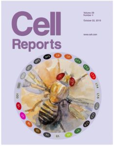
7. YCD Chen, SJ Park, RM Joseph, WW Ja and A Dahanukar.
Combinatorial pharyngeal taste coding for feeding avoidance in adult Drosophila. Cell Reports, October 22, 2019;29(4):961-973. doi: 10.1016/j.celrep.2019.09.036. [Cell Reports, Cover image, Oct. 22 2019 issue] [Link] [pdf] [Media]
Pharyngeal taste organs have been long assumed to as act a gatekeeper for monitoring food quality and controlling ingestion, but the functional evidence is lacking. Here we show how pharyngeal taste neurons drive the evaluation and feeding avoidance of different aversive tastants via a combinatorial sensory coding strategy. Notably, researchers have often overlooked at pharyngeal taste organs when interpreting results of feeding behavior experiments. Our findings not only make basic contributions to the fields of taste biology and regulation of insect feeding behavior but also have an impact on strategies for developing anti-feedants as part of integrated insect management strategies.
6. YCD Chen, S Ahmad, K Amin and A Dahanukar. A subset of brain neurons controls regurgitation in adult Drosophila melanogaster. Journal of Experimental Biology, October 1, 2019; 222 (Pt 19). doi: 10.1242/jeb.210724. [Link] [pdf]
Animals control food intake to meet their nutritional need and avoid toxic compounds in their environment. Such feeding control is tightly regulated through orchestrating the sensory-motor neuronal circuits. However, the neuronal circuits underlying individual feeding behaviors have yet to be elucidated. Here we screened ~200 GAL4 lines for neurons controlling feeding behaviors and found one line (VT041723-GAL4) that labels neurons receiving pharyngeal taste input and controlling regurgitation. This study act as a starting point for dissecting the regurgitation circuits, and a better understanding of feeding neuronal circuits will provide a novel design in controlling insect pests.
5. YCD Chen, SJ Park, WW Ja and A Dahanukar. Using Pox-neuro (Poxn) mutants in Drosophila gustation research: a double-edged sword. Frontiers in Cellular Neuroscience, October 24, 2018; 12(382). doi: 10.3389/fncel.2018.00382. [Link] [pdf]
Pox-neuro (Poxn) mutants have been widely used in Drosophila gustation research. In these mutants, all external taste hairs are transformed into mechanosensory hairs, offering a tool with which to identify taste-enriched genes and investigate behavioral consequences of ablating taste input. However, recent work indicates that chemoreceptor expression and function are likely intact in internal pharyngeal taste organs in Poxn mutants. Here, we present the advantages and shortcomings of using Poxn mutants in gustation research and reevaluate some of the established conclusions derived from previous work with these mutants. We also discuss the use of Poxn mutants for studying pharyngeal taste.
4. YCD Chen and A Dahanukar. Molecular and cellular organization of taste neurons in adult Drosophila pharynx. Cell Reports, December 5, 2017; 21(10):2978-2991. doi: 10.1016/j.celrep.2017.11.041. [Link] [pdf] [Media]
In Drosophila, most gustation research has focused on neurons residing in external taste hairs in the labellum, legs, and wing margins. There remains a complex repertoire of pharyngeal taste neurons lying in an anatomical position to control food intake, which has been largely overlooked. In this study, I carried out a large-scale, systematic analysis of the molecular organization of pharyngeal taste neurons, and established a detailed receptor-to-neuron map for all pharyngeal taste neurons. The detailed map contributes to the development of a minimal taste system model where only pharyngeal GRNs are being manipulated and to the study of the roles of each pharyngeal GRNs in feeding control.
3. EE LeDue, YC Chen, AY Jung, A Dahanukar and MD Gordon. Pharyngeal sense organs drive robust sugar consumption in Drosophila, Nature Communications, March 25, 2015; 6:6667. doi: 10.1038/ncomms7667. [Link] [pdf]
Much is known about sugar sensing through peripheral sweet taste neurons in the fly’s legs and labellum. However, sugar sensing by the pharyngeal taste organs has remained unexplored due to their inaccessibility to electrophysiology and the lack of genetic tools to specifically manipulate their function. Here, we characterize the physiological and behavioral roles of pharyngeal sweet taste neurons and identify a subset of gustatory receptor genes that are expressed in these neurons. We also show that mutant flies lacking all external taste hairs are nevertheless capable of selecting sugars by virtue of pharyngeal taste neurons, providing direct functional evidence of pharyngeal taste neurons.
2. PC Huang, YT Hsiao, SY Kao, CF Chen, YC Chen, CW Chiang, CF Lee, JC Lu, Yijuang Chern and CT Wang. Adenosine A2A Receptor Up-regulates Retinal Wave Frequency via Starburst Amacrine Cells in the Developing Rat Retina, PLoS ONE, April 28, 2014; 9(4):e95090. doi: 10.1371/journal.pone.0095090. [Link] [pdf]
Following our previous Syt I work, we further show that adenosine receptor is expressed in wave-initiating neurons, and adenosine signaling up-regulates the frequency of retinal waves. Retinal waves propagate through the developing retina, inducing similar burst patterns in the thalamus and visual cortex to form sensory loops. The etiology of schizophrenia and autism has been attributed to a developmental disorder of neural connectivity in the formation of sensory maps. Thus, identifying the roles of exocytotic or neuromodulator molecules that regulate the properties of retinal waves in developing neural circuits would help developing novel therapeutic methods for human psychiatric diseases.
1. CW Chiang, YC Chen, JC Lu, YT Hsiao, CW Chang, PC Huang, YT Chang, PY Chang and CT Wang. Synaptotagmin I Regulates Patterned Spontaneous Activity in the Developing Rat Retina via Calcium Binding to the C2AB Domains, PLoS ONE, October 16, 2012; 7(10): e47465. doi: 10.1371/journal.pone.0047465. [Link] [pdf]
During a developmental critical period, the spontaneous bursts of action potentials fire periodically and propagate across a population of developing retinal neurons to refine the visual circuits, termed as “retinal waves”. However, how retinal waves occur and how they are regulated remain mysterious. Here we show that the firing patterns of retinal waves can be modulated by a calcium sensor protein (Synaptotagmin I, Syt I) expressing in the wave-initiating neurons. We also show that the two calcium-binding domains of Syt I regulate the temporal properties of retinal waves during development. In this study, we also developed an in vitro electroporation method for gene delivery into retinas.
Peer-Reviewed Commentaries
5. V Menon* and YCD Chen*. Commentary: The role of the anion in salt (NaCl) detection by mouse taste buds. Frontiers in Cellular Neuroscience, November 8, 2019; 13(502). doi: 10.3389/fncel.2019.00502. *equal contribution. [Link] [pdf]
This commentary discusses a Journal of Neuroscience article published by Dr. Nirupa Chaudhari’s group. We provide a short overview of the topic and questions addressed in the commented paper, a critical review of the key findings, and a brief summary of the significance of the paper. We place this in the context of a broader picture of diverse cellular and molecular mechanisms of salt taste transduction among different taste cell types. We raise an interesting parallel with Drosophila sweet-sensing neurons in responding to high salt that could have some interesting implications for taste coding. We also point out the potential inhibitory role of K+ in salt taste transduction.
4. YCD Chen and A Dahanukar. DH44 neurons: Gut-brain amino acid sensors. Cell Research, October 11, 2018; 0:1–2. doi: 10.1038/s41422-018-0101-z. [Link] [pdf]
This research highlight discusses a Cell Research article published by Dr. Liming Wang’s group. We summarize the known cellular and molecular pathways involved in sensing amino acids in Drosophila and point out the diverse role of DH44 neuroendocrine cells in the Drosophila brain in sensing both sugars and amino acids. We propose a compelling brain-gut microcircuit that senses sugars and amino acids and promotes food intake through DH44 signaling and raising several interesting questions about the cellular and molecular mechanisms integrating information about both sugars and amino acids for balancing nutrient intake with metabolic needs.
3. J Clark* and YCD Chen*. Phosphorylation Switch of Orco Shapes the Sense of Smell in Insects. The Journal of Neuroscience, January 31, 2018; 38(5):1058-1060. doi: 10.1523/JNEUROSCI.3157-17.2017. *equal contribution. [Link] [pdf]
This commentary discusses a Journal of Neuroscience article published by Dr. Dean Smith’s group. We provide a short overview of the olfactory adaptation and performed a critical analysis of the commented papers. We raise a model of how olfactory responses by odorant coreceptor (Orco) phosphorylation are modulated over time. Given that a screening system for small molecules that target phosphorylation-dependent protein–protein interaction has been established, we also propose modulation of olfactory responses by the phosphorylation status of Orco could lead to the discovery of new generations of insect repellents against insect vectors and agricultural pests.
2. YCD Chen. Commentary: Retinal Waves Modulate an Intraretinal Circuit of Intrinsically Photosensitive Retinal Ganglion Cells. Frontiers in Neural Circuits, January 8, 2018; 11(113). doi: 10.3389/fncir.2017.00113. [Link] [pdf]
This commentary discusses a Journal of Neuroscience article published by Dr. Marla Feller’s group. I summarize the mechanisms underlying the activity-dependent modulation of intrinsically photosensitive retinal ganglion cells (ipRGCs) gap junction networks in the developing retina. I raise several interesting future directions, such as identifying subtypes of ipRGCs, characterizing their light responses, and determining which subtypes are coupled and which are not. I also place it in the context of other recent work in the visual cortex and propose that the activity-dependent regulation of gap junction coupling networks might be a conserved feature in the developing nervous systems.
1. YC Chen. The interactions between bitter and sweet taste processing in Drosophila, The Journal of Neuroscience, July 1, 2015; 35(26):9542-9543. doi: 10.1523/JNEUROSCI.1552-15.2015. [Link] [pdf]
This commentary discusses a Journal of Neuroscience article published by Dr. Frédéric Marion-Poll’s group. To promote behavioral avoidance, bitter compounds have been proposed to not only activate bitter taste neurons but also inhibit sweet taste neurons. Here, I summarize different mechanisms that bitter compounds can mask the sweetness in food mixture in Drosophila. I raise future directions to study how previously identified GABAergic interneurons inhibit sweet taste neurons. Since the suppression of sweet taste by bitter compounds has been observed in other insects, a better understanding of its underlying mechanism could be used for designing novel feeding deterrents for insect pests.